
Fig. 2.
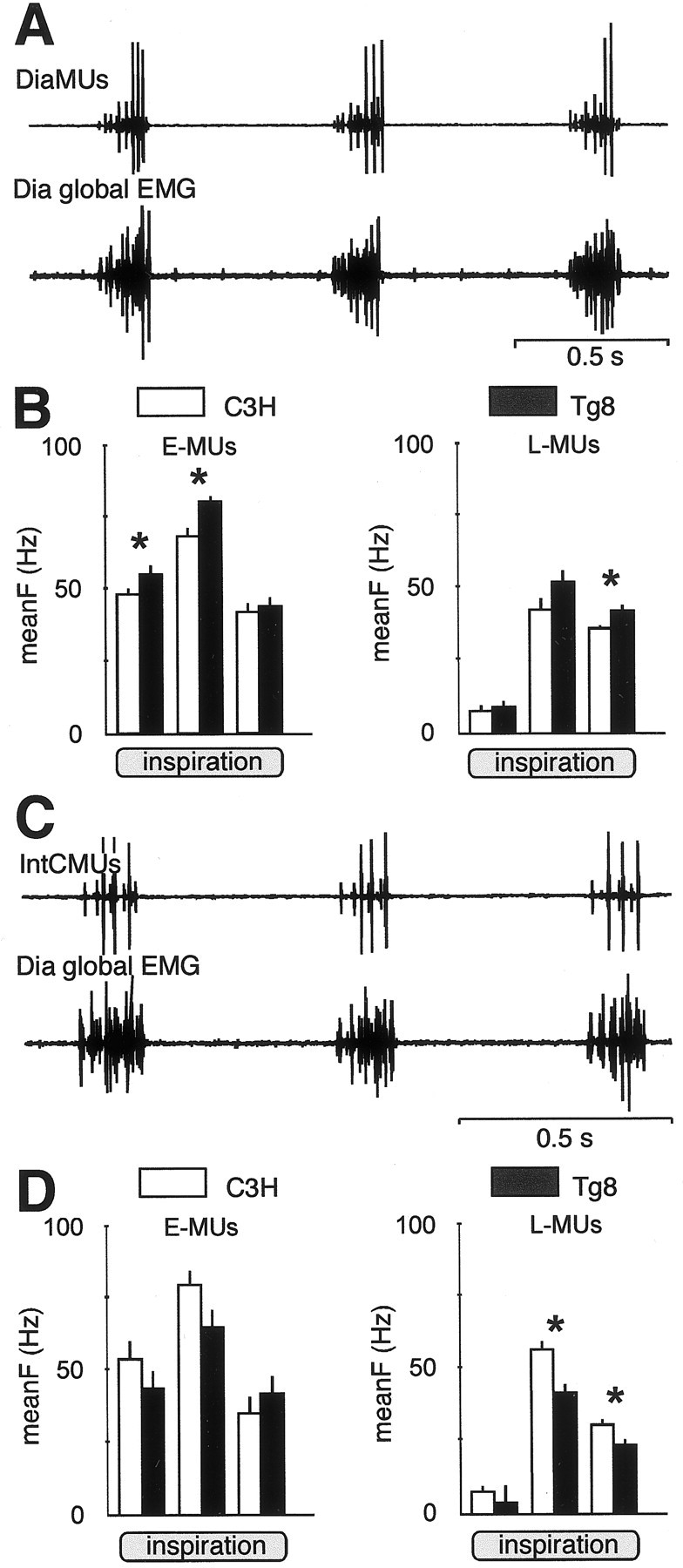
The firing activity of inspiratory motor units is different in Tg8 and C3H mice. A, Simultaneous EMG recordings from three motor units of the diaphragm (DiaMUs, top trace) and from theDia global EMG (bottom trace). The three DiaMUs are identified by their spike amplitudes. The small spikes correspond to an early-recruited MU (E-MU); the mid and large spikes correspond to two late-recruited MUs (L-MUs). B, DiaMUs are more active in Tg8 mice (black bars) than in C3H mice (white bars), as shown by the histograms displaying the mean firing frequency (meanF ± SEM, Hz) for E-MUs (left histograms) and L-MUs (right histograms) during the first, second, and last third of inspiration (* indicates a statistically significant difference at p < 0.05). In C3H mice, E-MUs = 16 and L-MUs = 20; in Tg8 mice, E-MUs = 18 and L-MUs = 18. C, Simultaneous EMG recordings from two intercostal motor units (IntCMUs,top trace) and from the Dia global EMG(bottom trace). D, The histograms of the meanF of the IntCMUs in Tg8 mice (black bars) and in C3H mice (white bars) show that E-MUs (left histograms) have similar firing activity during the three thirds of inspiration, whereas L-MUs (right histograms) are significantly more active during the last two-thirds of inspiration in the C3H mice (* indicates a statistically significant difference atp < 0.05). In C3H mice, E-MUs = 9 and L-MUs = 42; in Tg8 mice, E-MUs = 6 and L-MUs = 37.