
Fig. 3.
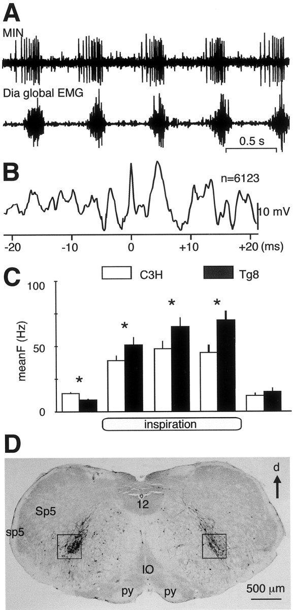
The medullary inspiratory neurons are more active in Tg8 than in C3H mice. A, Simultaneous recordings from a MIN discharge (top trace) and from the ipsilateral Dia global EMG (bottom trace) in a C3H mouse. B, Spike-triggered-averaging analysis of the Dia global EMG from spikes (n = 6123) occurring in the recorded MIN in A shows that the Dia activity increases 5 msec after the occurrences of MIN spikes. C, On the basis of recordings from 12 MINs in C3H mice (white bars) and 19 MINs in Tg8 mice (black bars), the histograms show the mean firing frequency (meanF ± SEM, Hz) of these neurons during respiration. MINs start to fire before inspiration, increase their firing activity during the three thirds of inspiration, and fire at a low rate during the beginning of expiration before becoming silent (* indicates a statistically significant difference at p < 0.05). D, Four days after the rabies virus has been injected into the left part of the Dia, labeled neurons were found in the medullary areas in which MINs have been recorded (black squares). d, Dorsal; IO, inferior olive; sp5, spinal trigeminal tract; Sp5, spinal trigeminal nucleus;py, pyramidal tract; 12, hypoglossal nucleus.