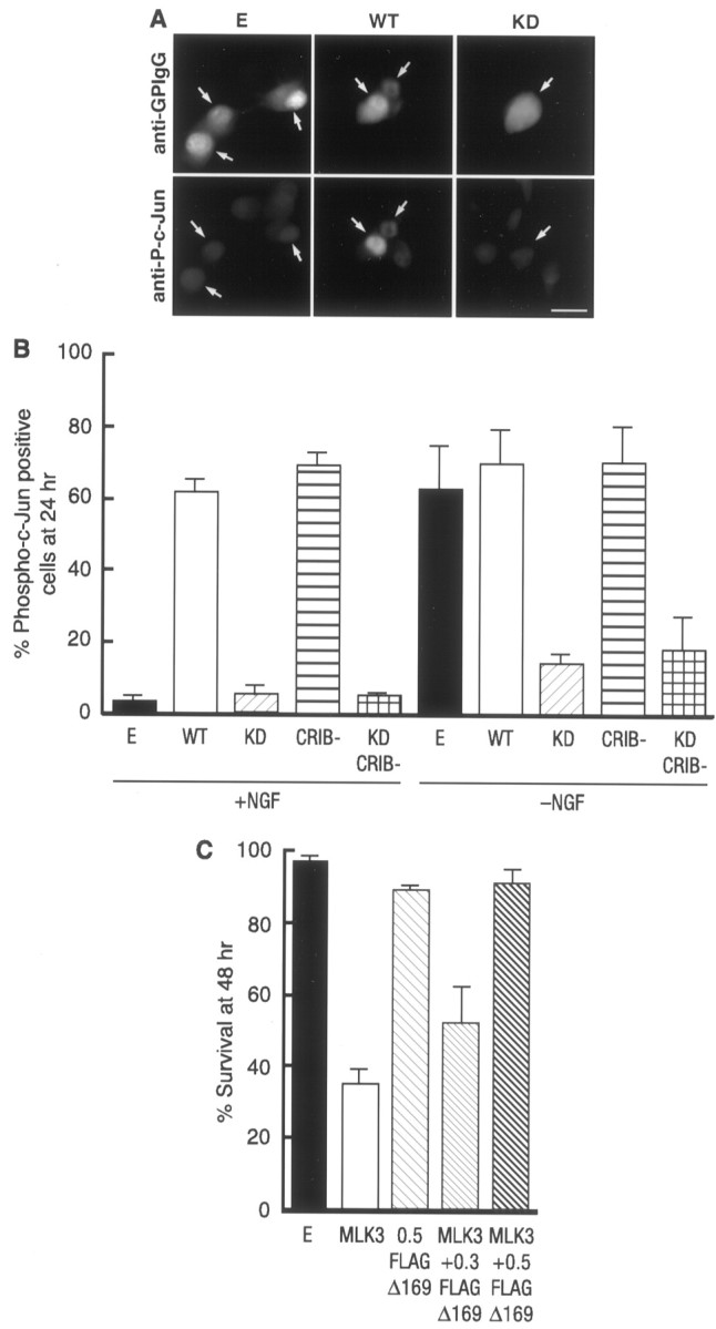
Fig. 6.

MLK3 activates the JNK pathway in neurons. Shown is the MLK3-dependent phosphorylation of c-Jun in SCG neurons. SCG neurons were microinjected either with 0.3 mg/ml of an empty expression vector or the different MLK3 mutants, together with 5 mg/ml guinea pig IgG to detect the injected cells, and maintained in the presence of NGF or withdrawn from NGF as indicated. After 24 hr the cells were fixed, permeabilized, and stained with an anti-guinea pig IgG antibody (A, top) and with a specific anti-phospho-c-Jun antibody (A, bottom). Only the cells in which phospho-c-Jun staining was clearly above background were scored as positive. Arrows indicate injected cells. Scale bar, 30 μm. The results were quantified and represented as a bar graph (B). They are the mean of three independent experiments ± SEM. C, FLAG-Δ169 blocks MLK3-induced apoptosis. FLAG-Δ169 at the indicated concentrations (in mg/ml) and 0.1 mg/ml of WT MLK3 were microinjected into 5- to 7-d-old SCG neurons and maintained in the presence of NGF. The percentage of surviving cells was assessed 48 hr later. The results are the mean of three independent experiments ± SEM.
