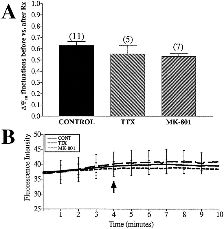
Fig. 6.
ΔΨm fluctuations are not attributable to synaptic activity. Untreated neurons were loaded with JC-1 and imaged for 4 min before the addition of drugs (arrow). TTX (200 nm) (a Na+ channel blocker) or 10 μm MK-801 (NMDA antagonist) were perfused over the cells for 5 min, and fluorescence intensity and number of fluctuations per minute per 1000 ROIs were calculated. A, The ratio of fluctuations occurring during the baseline to those occurring after drug treatment. This corrected for the consistent decrease in fluctuations observed in untreated mitochondria (see Fig. 3). Data are presented as means ± SEM, and the numbers above each barequal the number of fields imaged (∼1000 ROIs per field). Neither treatment had a significant impact on ΔΨm fluctuations (TTX, t = 1.03, df = 14, p> 0.05; MK-801, t = 1.98, df = 16,p > 0.05). B, Average fluorescence intensity of all mitochondria imaged, with representative error bars indicating SEM. Neither treatment altered basal fluorescence.

