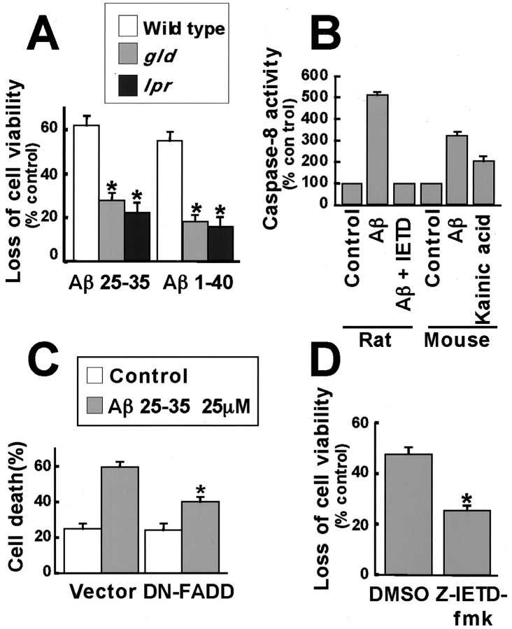
Fig. 8.
The Fas ligand–Fas pathway is required for Aβ-induced apoptosis. A, Cortical neurons were prepared from wild-type, gld, and lprmice embryos and treated with 25 μm Aβ25–35 for 24 hr or 25 μm Aβ1–40 for 72 hr. Cell viability was assessed by the MTS assay. Data represent mean ± SEM (n = 5). *p < 0.05, significantly different from wild type (ANOVA and Dunnett's test).B, Neurons from rat and mouse were treated with 25 μm Aβ25–35 and the caspase-8 inhibitor, Ζ-IETD-fmk, or with 200 μm kainic acid. Caspase-8 activity was assessed by a fluorometric assay kit. Data represent mean ± SEM (n = 6 for rats; n = 4 for mice). C, Cortical neurons were cotransfected with 4 μg of an empty vector or DN-FADD and 0.7 μg of CMV-LacZ and 0.3 μg of CMV-EGFP. After 2 d, the cells were treated with 25 μm Aβ25–35 for 24 hr. The cells were fixed and immunostained with anti-β-Gal antibody. Nuclei were stained with Hoechst 33258. Transfected cells were identified as those that expressed green fluorescent protein, and of these cells, apoptotic neurons were scored as those that display condensed and fragmented nuclei. Data represent mean ± SEM of four wells from a typical experiment. D, Neurons were pretreated with DMSO or 100 μm caspase 8 inhibitor Ζ-IETD-fmk for 1 hr, and then 25 μm Aβ25–35 was added, and apoptosis was determined by the MTS assay. Data represent mean ± SEM of seven wells from a typical experiment. *p < 0.05, significantly different from vector or control (ANOVA and Dunnett's test or Student's t test).

