Fig. 2.
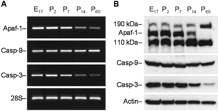
Analysis of age-dependent expression of Apaf-1, caspase-9, and caspase-3 mRNA and proteins in rat cortex.A, RT-PCR analysis of the abundance of transcripts encoding rat Apaf-1, caspase-9 (Casp-9), and caspase-3 (Casp-3) in cortex of E17 or P2, P7, P14, and P60 rat brains. Total RNA from rat cortex on the indicated days of development was subjected to RT-PCR with primers specific for Apaf-1, caspase-9, and caspase-3. Amplification of 28S rRNA was used as an internal control. The PCR products were analyzed by electrophoresis through an agarose gel and visualized after staining with ethidium bromide.B, Western blot analysis of the abundance of Apaf-1 and procaspases-9 and -3 in the protein extracts isolated from rat cortex on the indicated days of rat development. Eighty microgram aliquots of cytosolic protein extracts isolated from rat brain cortex at indicated developmental stages were subjected to 5% (Apaf-1) or 12% SDS-PAGE and transferred to a nitrocellulose filter. The filters were probed with a polyclonal anti-Apaf-1 antibody (AB16941; Chemicon), a monoclonal anti-caspase-9 antibody (clone 5B4; MBL), or a rabbit polyclonal antibody against caspase-3 (H-277, Santa Cruz Biotechnology). The antigen–antibody complexes were visualized by an ECL method as described in Materials and Methods. β-Actin protein abundance was used as an additional control for gel loading and transfer. These experiments were repeated four times with similar results.

