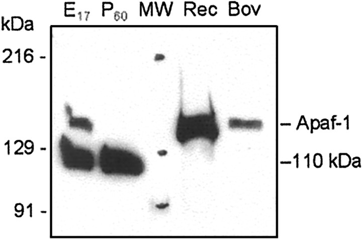
Fig. 3.
Analysis of anti-Apaf-1 antibody specificity. Fifty micrograms of cytosolic proteins from E17 and P60 rat cortex, 20 ng of recombinant human Apaf-1 (Rec), 10 ng of Apaf-1 purified from bovine thymus (Bov), and 5 μl of prestained standards (MW; Bio-Rad, catalog #161-0324) were separated in 5% SDS-PAGE followed by staining with a polyclonal antibody (AB16941; Chemicon). The preparation of purified bovine Apaf-1 demonstrated Apaf-1 activity in the in vitroreconstitution system with cytochrome c (data not shown). Results show the location of the Apaf-1 protein band above the extensively stained 110 kDa protein of unknown origin.

