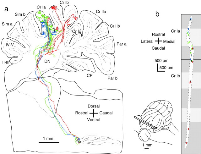
Fig. 4.
Climbing fiber distribution in a narrow longitudinal area in the hemisphere. a, Lateral view of trajectories of three olivocerebellar axons (colored) reconstructed on 31 serial coronal sections. A tracer injection was made in the rostral portion of the dorsal lamella of the principal subnucleus of the inferior olive. Of 23 labeled climbing fibers, 18 climbing fibers arose from these three axons, and the other five labeled climbing fibers (gray) from other olivary neurons. The outline of the cerebellum is deformed to show all climbing fibers. b, The distribution of climbing fibers was plotted on the unfolded hemisphere along a tilted parasagittal plane.Inset shows the tilted parasagittal plane in which climbing fibers were aligned. Colors used for the climbing fibers inb correspond to those used for individual axons ina. Light and dark grayareas in the unfolded scheme represent the cerebellar cortex exposed in the cerebellar surface and hidden in the sulci, respectively.Dotted line indicates the contour of the distribution area.

