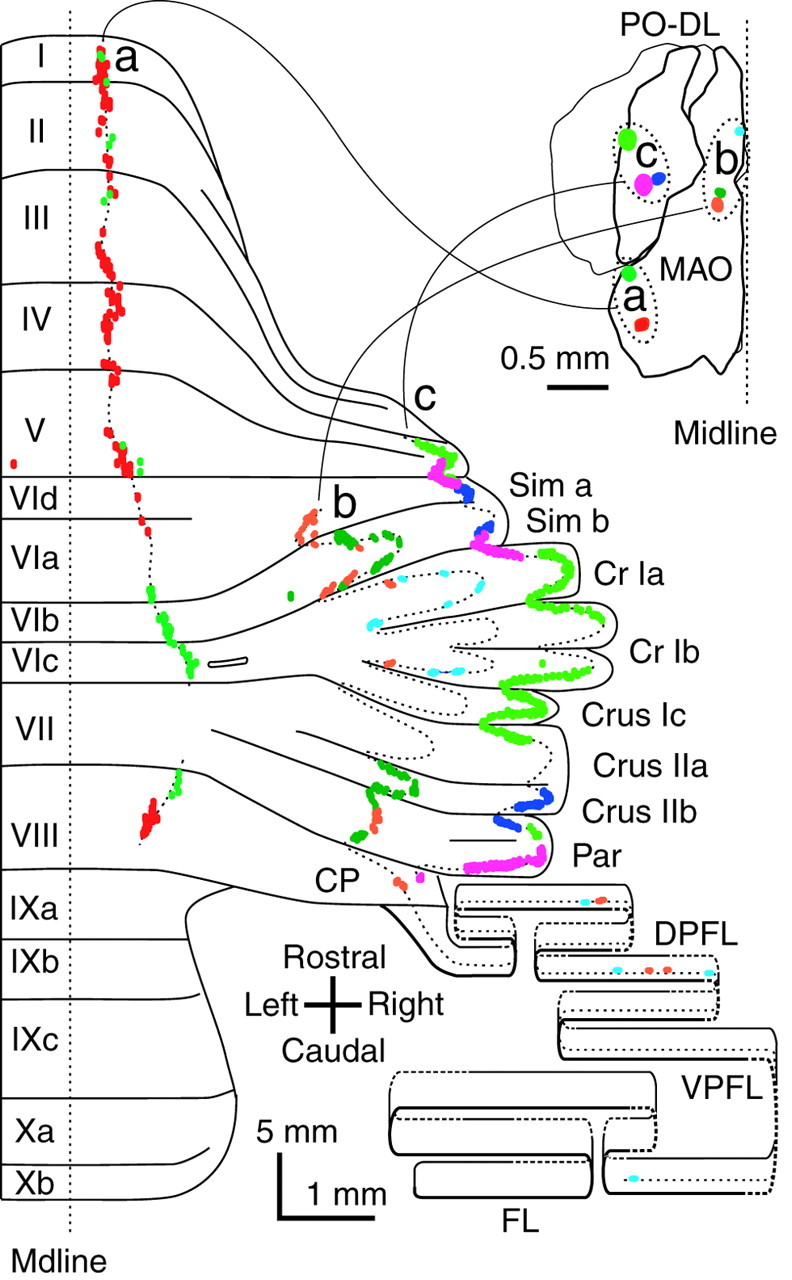
Fig. 5.

Projection patterns of climbing fibers originating from adjacent areas in the inferior olive showing complementary segments within the same longitudinal band. Climbing fibers labeled by small injections in two adjacent sites in the lateral and caudal portions of the MAO (green andred) (a), three sites in the medial and rostral portions of the MAO (orange, dark green, and light blue) (b), and three sites in the lateral portion of the dorsal lamella of the principal olive (PO–DL) (yellow–green, pink, andblue) c, Results of eight experiments are presented in three groups based on olivary injection sites and their related cerebellar projections (a, vermis;b, medial hemisphere or pars intermedia;c, lateral hemisphere). Inset shows the dorsal view of the injection sites (colored spots) in the inferior olive. Dotted lines drawn along labeled climbing fibers in the cerebellar cortex indicate putative single longitudinal bands. Each colored dot, which is often fused to others, indicates a labeled climbing fiber.
