
Fig. 6.
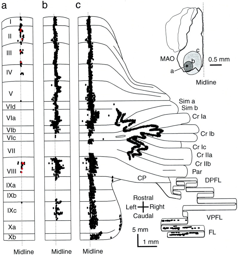
The distribution patterns of climbing fibers labeled by injections of different volumes of tracer into the inferior olive indicating substructures in the zonal organization of the olivocerebellar projection. a, Two longitudinal segments in lobules I–IV and VIII in the vermis along the midline produced by a small injection in the caudal portion of the MAO (right inset). Red dots indicate climbing fibers of a single reconstructed olivocerebellar axon terminating in both areas. b, Longitudinal band-shaped pattern occupying most of the lobules in the vermis along the midline produced by a medium-sized injection including the site in a.c, Striped pattern produced by an injection larger than in b and covering the sites ina and b in the MAO. Curvatures of some lateral bands are attributable to the tilt of the longitudinal plane and the foliation of the cortex. Each dot, which is often fused to others, indicates a labeled climbing fiber.