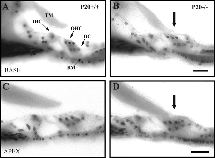
Fig. 2.
Altered morphology of the OC tectorial membrane in Igf-1−/− P20 mice. Nissl staining of celloidin-embedded cross sections of the OC of P20Igf-1+/+ (A, C) andIgf-1−/− (B, D) animals. A and B show basal turns, whereas C and D show apical turns of the cochlea. Physical attachment of the tectorial membrane to the hair cells was noticed in all sections of P20Igf-1−/− (B, D, arrows). BM, Basilar membrane;DC, Deiters' cells; IHC, inner hair cells; OHC, outer hair cells; TM,tectorial membrane. Scale bars, 30 μm.

