Fig. 3.

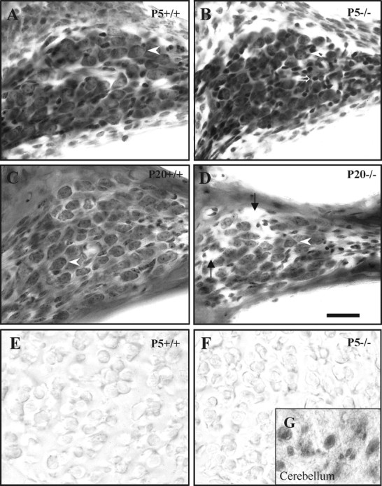
Altered cochlear ganglion morphology inIgf-1−/− mice. Nissl staining of the ganglion in the cochlear basal turn inIgf-1+/+ (A, C) andIgf-1−/− (B, D) mice at P5 (A, B) and P20 (C, D). At P5, the cochlear ganglion and its neurons show similar morphology in both genotypes (arrowheads), except thatIgf-1−/− mouse also show a subtype of abnormally small, strongly chromaffinic cells (open arrow). At P20, ganglion cells are noticeably reduced both in size and number in Igf-1−/− mice. This reduction, despite the presence of enlarged intercellular spaces (arrows), leads to a considerable decrease in the ganglion cross-sectional area. E and Fshow negative PCNA expression in the cochlear ganglion cells of P5Igf-1+/+ (E) and Igf-1−/−(F) mice. The inset(G) shows a positive control of PCNA-positive cerebellum cells from the same section of P20Igf-1−/− mouse shown inF. Scale bar, 30 μm.
