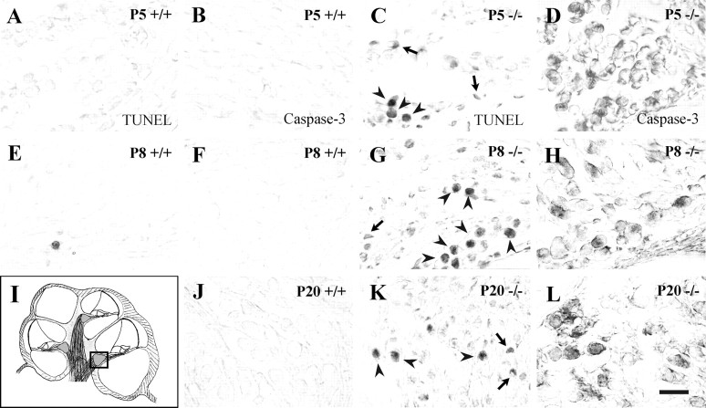
Fig. 5.
Apoptotic cell death in the cochlear ganglion ofIgf-1−/− mice. TUNEL labeling (A, C, E, G, K) and detection of activated caspase-3 expression (B, D, F, H, J, L) were performed on paraffin sections from normal (Igf-1+/+) and mutant (Igf-1−/−) mice at postnatal days 5, 8, and 20. The area analyzed is shown in a schematic drawing of the cochlea in which the square indicates basal turn cochlear ganglia (I). Note the increase in apoptotic nuclei (C, G, K) and intense activated caspase-3 immunostaining (D, H, L) in the mutant mice.Arrowheads point to apoptotic neurons, whereasarrows point to dying glial cells. The sections correspond to basal turns of the cochlea. Scale bar, 30 μm.

