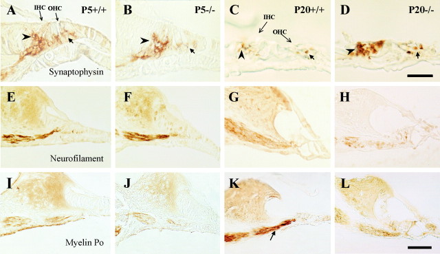
Fig. 7.
Differential expression of neural and glial markers in the innervation of the organ of Corti. Immunohistochemical staining of midmodiolar paraffin sections of basal turns of the cochlea with synaptophysin (A–D), neurofilament 200 kDa (E–H), and myelin P0(I–L) antibodies. Left panels(A, E, I) correspond to P5Igf-1+/+ samples, middle-left panels (B, F, J) to P5Igf-1−/−, middle-right panels (C, G, K) to P20Igf-1+/+, and right panels (D, H, L) to P20Igf-1−/− mice. At P5 (A, B), synaptophysin immunoreactivity appears diffuse and displays a “cup-like” shape surrounding the IHCs (arrowheads) and OHCs (arrows). At P20 (C, D), the synaptophysin staining pattern is better defined at the base of IHC and nerve fibers in the Igf-1+/+ controls than in Igf-1−/− mutants. NF-200K and myelin P0 immunostainings do not evidence major differences at P5 but are clearly less intense in P20Igf-1−/− mice (H, L) than in Igf-1+/+ controls (G, K, arrow). Scale bars: A–D, 20 μm;E–L, 30 μm.

