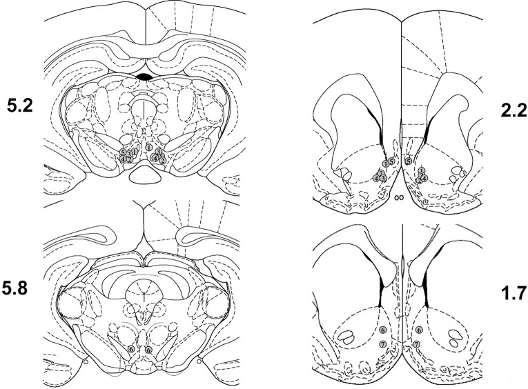
Fig. 1.
Histological verification of infusion sites. The location of infusions is shown for all rats that received intra-VTA infusions of M100907 (left panels) and intra-NAc infusions of RS 102221 (right panels). Plates are taken fromPaxinos and Watson (1997), and the numbers beside each plate correspond to millimeters from bregma.

