
Fig. 1.
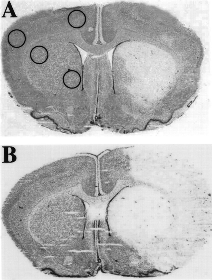
Cresyl violet-stained coronal brain sections from estrogen-treated (A) and untreated (B) ovariectomized female rats after stroke. The area of pallor delineates the “core” of cerebral infarction, and the area immediately surrounding that core represents the “penumbra” of ischemic injury. Estrogen treatment resulted in 40% reduction in cerebral infarct (n = 4 per group).Circles delineate areas from which tissue micropunches were extracted for bcl-2 mRNA quantification using RNase protection assay.