
Fig. 2.
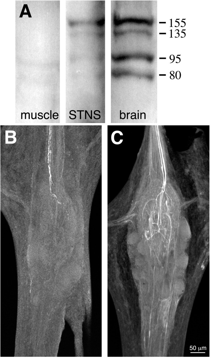
NOS expression and activity in the STNS.A, Western blot of proteins extracted from crab muscle, STNS, and brain using a polyclonal antibody raised against a conserved domain of NOS (uNOS). Molecular weights (in kilodaltons) are indicated to the right. B,C, Whole-mount confocal projections of stomatogastric ganglia reacted with a polyclonal antibody raised against citrulline, a byproduct of NOS enzymatic activity. Each projection is a complete image through the ganglion. B, Putative NOS enzymatic activity is present under basal conditions in two projection neurons that enter the STG via the stn (at top;n = 5). Citrulline accumulation is localized exclusively to the posterior stn and the STG (data not shown). C, Citrulline staining in the two input fibers is enhanced when the perineural sheath is removed and the STG is preincubated for 30 min in 1 mml-arginine (n = 3). Note extensive branching of citrulline-containing terminals in the central synaptic region of the ganglion (neuropil). Because the citrulline antibody cross-reacts with high concentrations of arginine in the nervous system (Eliasson et al., 1997), the faint background staining in unidentified axons may reflect arginine accumulation in these processes.