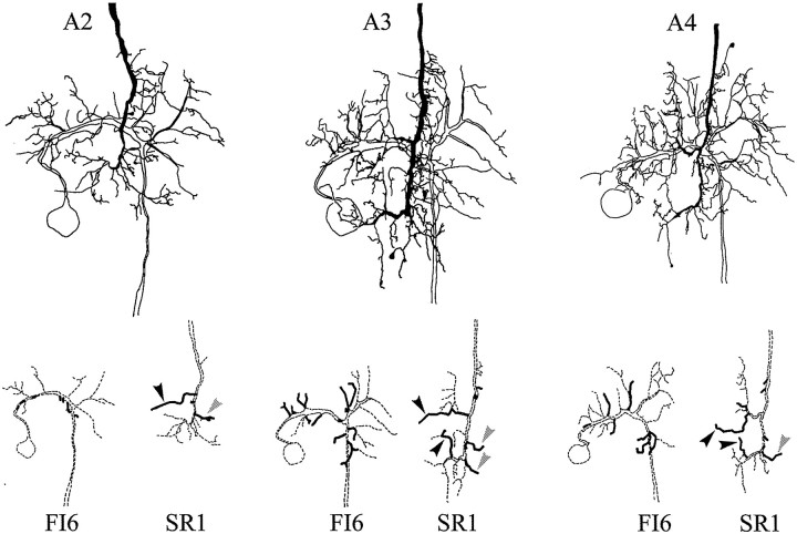
Fig. 8.
Camera lucida drawings of paired fills of an FI6 neuron and an SR1 axon from contralateral A2, A3, or A4. The primary structure of each neuron is drawn with dashed lines at the bottom; branches that make putative synaptic contacts are drawn with solid lines. Black arrowheads mark branches that contacted the main dendritic region of FI6; gray arrowheads mark branches that contacted FI6 near its spike-initiating zone (see Results).

