Fig. 4.
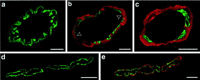
Cerebrovascular amyloid leads to smooth muscle cell loss. Confocal microscopy of double-immunolabeled vessels (green, smooth muscle actin; red, amyloid) in APP23 mice. a, Leptomeningeal vessel in an 8-month-old mouse shows no amyloid deposition and a complete layer of smooth muscle cells. b, Leptomeningeal vessel in a 19-month-old mouse shows focal disappearance of smooth muscle cells at the site of cerebrovascular amyloid (arrowheads).c, In 27-month-old mice, smooth muscle cells have greatly disappeared, and a thick sheet of amyloid covers the wall of a leptomeningeal vessel. d, e, Parenchyma in the neocortex of a 19-month-old mouse showing an unaffected (d) and an amyloid-laden vessel (e) in close anatomical proximity. Shown are superpositions of 0.9- to 5-μm-thick optical sections. Scale bars:a, 10 μm; b–e, 20 μm.

