
Fig. 7.
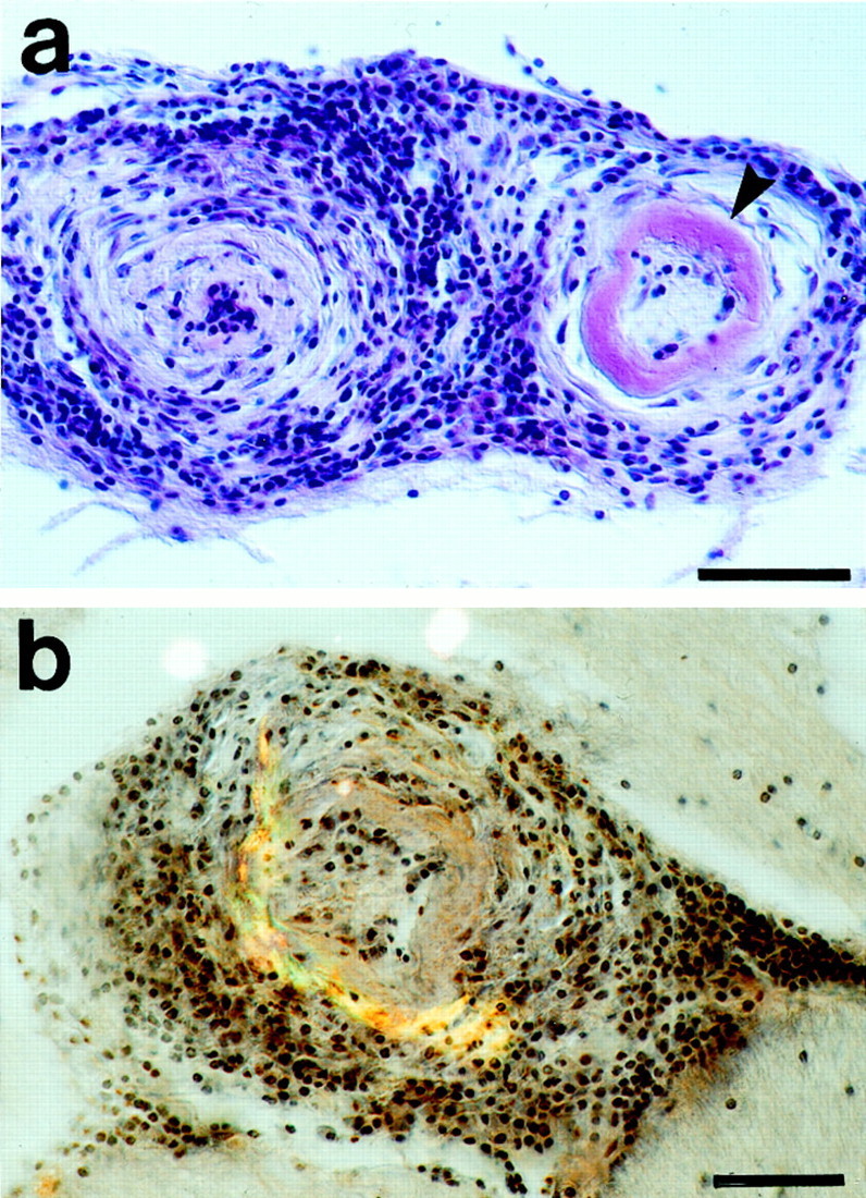
Vasculitis in aged APP23 mice with severe CAA.a, H&E staining of two vessels affected by a chronic lymphocytic vasculitis. Lymphocytic infiltrates are seen throughout the entire vessel walls. The vessel wall on the left appears thickened and the lumen is obliterated. There is severe amyloid deposition in the right vessel wall (arrowhead). b, Double-staining for H&E and for Congo red (green-yellow birefringence) reveals amyloid deposits in a vessel heavily affected by a lymphocytic vasculitis. Scale bars, 50 μm.