
Fig. 7.
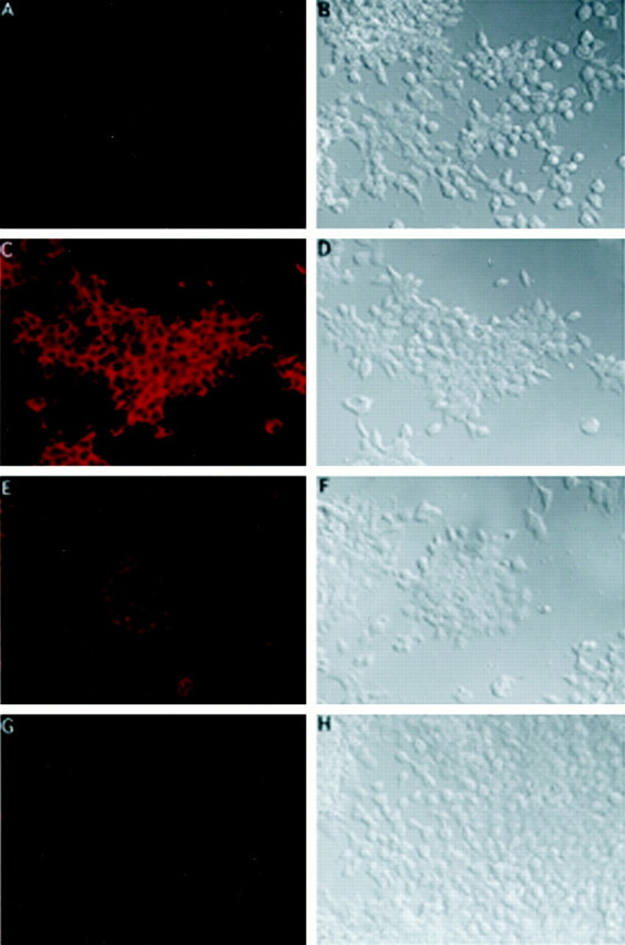
Immunohistochemical analyses of KOR protein expression in P19 cells. P19 stem cells (C,D) and cultures induced with RA for 3 (E,F) and 5 (G,H) d were stained with an anti-KOR antibody (Chen et al., 1999), followed by reaction with a Cy3-conjugated secondary antibody (C, E, G).D, F, and H show the bright-field image of C, E, andG, respectively. A negative control of P19 stem cells stained with preimmune serum is shown in A.B shows the bright-field image ofA.