Fig. 1.
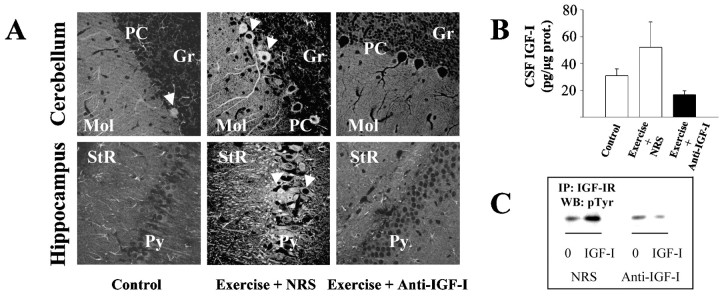
Infusion of an anti-IGF-I antiserum inhibits exercise-induced brain uptake of circulating IGF-I. A,Control sedentary animals show very low IGF-I labeling in the brain. Rats running in a treadmill for 15 d and receiving a simultaneous infusion of a nonblocking normal rabbit serum (NRS) show marked increases in IGF-I labeling throughout the brain. However, animals undergoing the same exercise schedule but receiving an infusion of an anti-IGF-I antiserum do not show increased brain IGF-I labeling. The cerebellar cortex and the hippocampus are shown as representative areas accumulating IGF-I after exercise (see references for further details). Arrows indicate typically labeled neurons.Mol, Cerebellar molecular layer; PC,Purkinje cell layer; Gr, cerebellar granule cell layer;Py, hippocampal pyramidal cell layer;StR, hippocampal stratum radiatum. B,Levels of IGF-I in the CSF of exercising animals receiving an infusion of NRS are 68% higher than in control, sedentary animals. However, exercising rats receiving an anti-IGF-I infusion do not show increased CSF IGF-I levels; n = 3 per group.C, The anti-IGF-I antibody used for blocking uptake of IGF-I blocks interaction of IGF-I with its receptor. The presence of the anti-IGF-I antiserum (20% in culture medium) completely blocks IGF-I-induced receptor tyr-phosphorylation in cerebellar neuronal cultures (right). When 20% NRS was used instead, IGF-I receptor was tyr-phosphorylated in response to IGF-I (left), indicating normal biological activity of IGF-I. Cerebellar cultures were lysed 3 min after addition of 100 nm IGF-I, immunoprecipitated (IP) with a polyclonal anti-IGF-I receptor antibody, and blotted (WB) with a monoclonal anti-pTyr antibody.

