
Fig. 2.
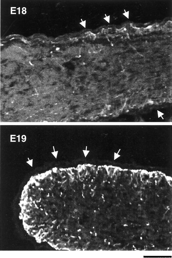
GFAP staining in developing optic nerves. Longitudinal cryosections of E18 and E19 optic nerves were stained by an anti-GFAP antibody and observed under higher magnification than that shown in Figure 1. The majority of GFAP staining at E18 is near the surface of the nerve, right underneath the pia (arrows). By E19, the staining beneath the pia has become much more intense and is beginning to extend more deeply into the nerve. Scale bar, 50 μm.