
Fig. 3.
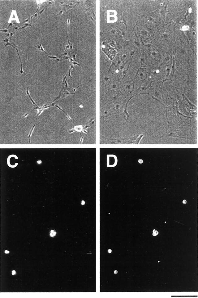
Morphology and immunoreactivity of purified VECs in culture. Phase-contrast micrographs of purified VECs (A) and pial cells (B) after 3 d in serum-free culture. VECs tend to be spindle-shaped (A), whereas the pial cells display a flat sheet-like appearance (B). To confirm the purity of the VECs, they were immunostained immediately after isolation with an anti-Tie2 antibody (C, D). All of the purified cells, indicated in (C) with the 4′,6′-diamidino-2-phenylindole (DAPI) nuclear stain, were Tie2+ (D). Scale bar, 100 μm.