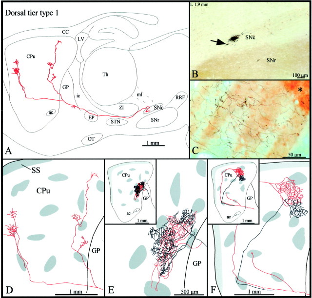
Fig. 2.
A, Camera lucida drawing of a dorsal tier type 1 SNc axon, as viewed in the sagittal plane. The compartmental organization of its terminal field is illustrated inD. B, Photomicrograph illustrating the BDA deposit in the dorsal tier of SNc. Note the CB+ cell bodies scattered in the dorsal tier and the CB+ neuropil that characterizes SNr but is absent in SNc. The arrow points to the neuron the axon of which is drawn in A. C, Photomicrograph showing part of the terminal arborization in the matrix of the axon drawn in E. The asteriskindicates one μR+ striosome. D–F, Striatal branching of three different type 1 axons. The shaded areas in the caudate-putamen indicate the μR+ striosomes and subcallosal streak.ac, Anterior commissure; CC, corpus callosum;CPu, caudate-putamen; LV, lateral ventricle;ic, internal capsule; OT, optic tract;SS, subcallosal streak; Th, thalamus. Definitions of abbreviations apply to all figures.

