
Fig. 3.
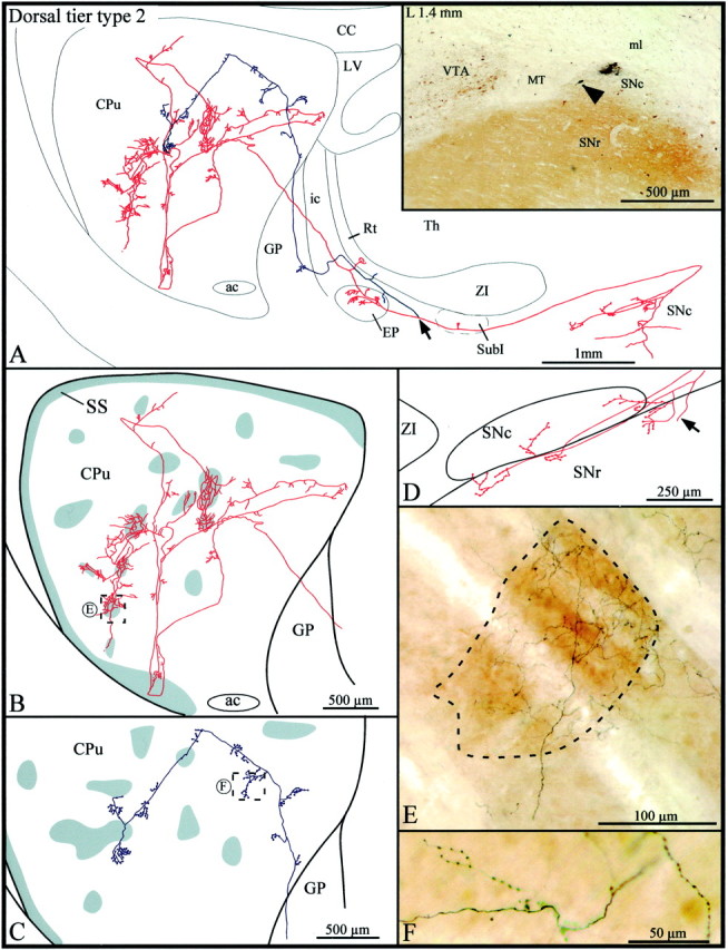
A, Camera lucida drawing of a dorsal tier type 2 SNc axon, the parent cell body of which is pointed out by the arrowhead in theinset. This neuron is located beside a very small injection site in the medial aspect of the dorsal tier of the SNc. Thearrow in the drawing indicates the level at which the main axon bifurcates, and the two axonal branches are represented inred and blue, respectively.B, C, High-power views of the striatal arborization of each axonal branch. The shaded areasindicate the μR+ striosomes and subcallosal streak. D, Camera lucida drawing showing the distribution of the local axonal collaterals in the substantia nigra. The arrow points to the main axon. E, F, Photographic enlargements of the terminal arborizations of each axonal branch at the level indicated by the dotted rectangles inB and C. SubI, Subincertal nucleus. Definition applies to all figures.