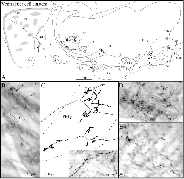
Fig. 8.
A, Camera lucida drawing of another ventral tier axon the neuron of which lies in a cell cluster located in the SNr, as viewed in the sagittal plane. The main axon (4) and its three main collaterals (1-3) are numbered to facilitate their identification. B, Photomicrograph of one axon collateral within the GP giving rise to several pedunculated terminal boutons. C, High-power view of the terminal arborization within the pedunculopontine tegmental nucleus (PPTg). The inset in C is a photographic enlargement of some terminal collaterals in the PPTg at the level indicated by the dotted rectangle in thedrawing in C. D,E, Photomicrographs of the terminal field within the paracentral thalamic nucleus (PC) (D) and of some axonal collaterals in the ventrolateral thalamic nucleus (VL) (E). FF, Fields of Forel;LPB, lateral parabrachial nucleus; MPB, medial parabrachial nucleus; PnO, pontine reticular nucleus, oral part; scp, superior cerebellar peduncle; VM, ventromedial thalamic nucleus. Definitions of abbreviations apply to all figures.

