
Fig. 9.
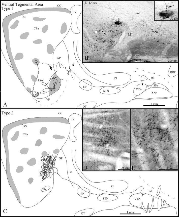
A, Sagittal representation of a type 1 VTA axon, with gray zones representing μR+ striosomes and subcallosal streak. The arrow points to the axon collateral that innervates a dorsal striosome before arborizing in the FStr. B, Photomicrograph illustrating the BDA deposit in the dorsal aspect of the VTA. The inset in B is a photographic enlargement of the injection site, and thearrow points to the axon that is drawn inA as it emerges from a primary dendrite.C, Sagittal representation of the axonal trajectory and striatal arborization of a type 2 VTA neuron. D, Photomicrograph of the dense terminal arborization in the matrix of the axon that is drawn in C. E, High-power view of some terminal fibers of the same axon at the striatal level. Definitions of abbreviations apply to all figures.