
Fig. 2.
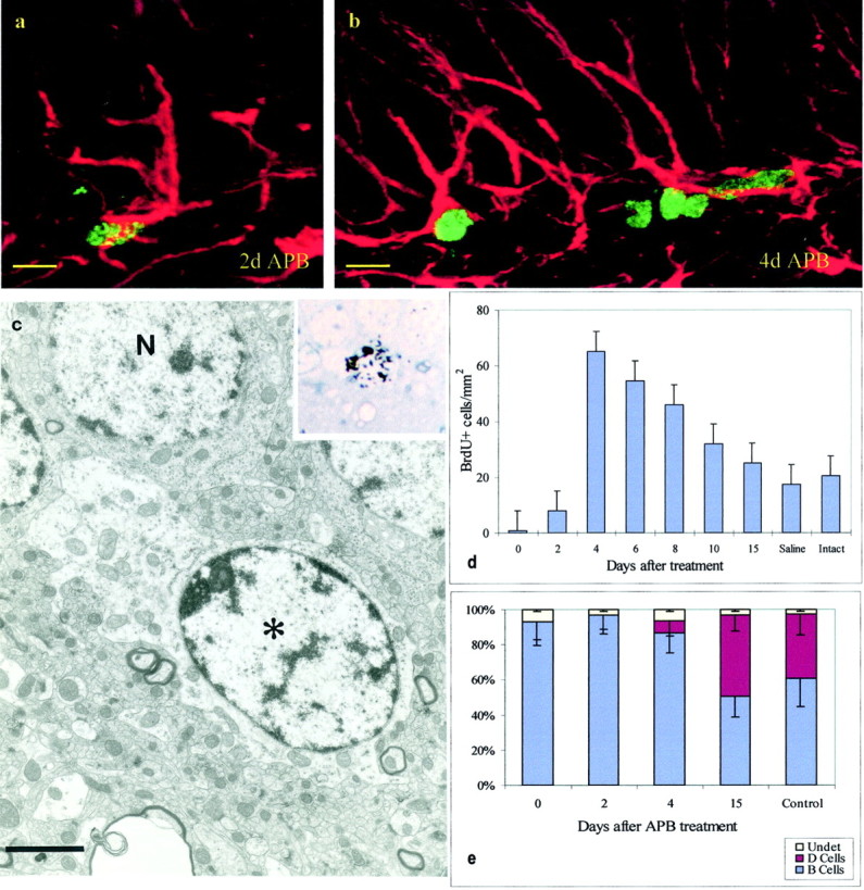
Cell reappearance after APB treatment.a, Cell labeled with BrdU (green) and GFAP (red) 2 d after termination of APB.b, Four days after APB, BrdU–GFAP-labeled cells remained, but some GFAP-negative BrdU-labeled cells were also present.c, Ultrastructural analysis of a [3H]-labeled cell (see autoradiogram in theinset) 2 hr after [3H]thymidine injection and 2 d after termination of APB treatment. This cell corresponds to a B cell. Note the light cytoplasm and irregular contours of the plasma membrane. d, Time course of reappearance of BrdU-labeled cells after termination of APB treatment;n = 5; mean ± SD. e, Cellular composition (EM) of the SGL at multiple survivals after APB termination; n = 3; 250 cells per animal; mean ± SD. Scale bars: a, b, 8 μm;c, 2 μm.