
Fig. 3.
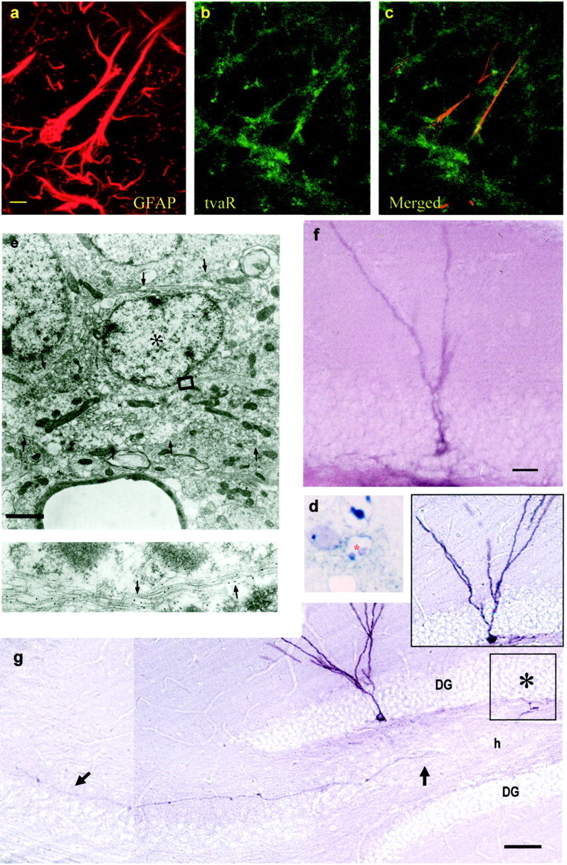
Retrovirally labeled SGL astrocytes give rise to new neurons in the granule cell layer. Double staining for GFAP and tvaR in the SGL of normal Gtva mice (a–c).a, Staining for GFAP. b, Staining for the tvaR. c, Merged fields showing combined immunostaining for GFAP and the tvaR. AP-positive cells identified after injection of RCAS-AP virus into the dentate gyrus of Gtva mice in eand d 2 d after viral injection cells were analyzed at the electron microscope. d, Plastic section (1.5 μm) of an AP-positive cell (asterisk) stained with NBT-BCIP (purple deposit; bright field). The cell was reembedded, resectioned, and stained with gold-conjugated antibodies to GFAP for ultrastructural analysis (e). Labeled cell corresponded to a B cell that contained intermediate filaments decorated by gold particles (arrows in the inset below). Thearrowheads in e show the irregular contours of the plasma membrane of this cell. f, Immature granule neurons start appearing 8 d after viral injection. g, Thirty days after RCAS-AP injection into SGL of Gtva mice, AP-labeled granule neurons are present in the dentate gyrus. These cells have the characteristic dendritic arbors in the molecular layer and an axon (arrows) that projects to CA3. The cell in the inset gives rise to the mossy fiber shown in g. This cell body is in the adjoining section in the approximate location shown by the asterisk. Scale bars: a, 5 μm (for a–c);e, 2 μm; f, 10 μm; g, 100 μm.