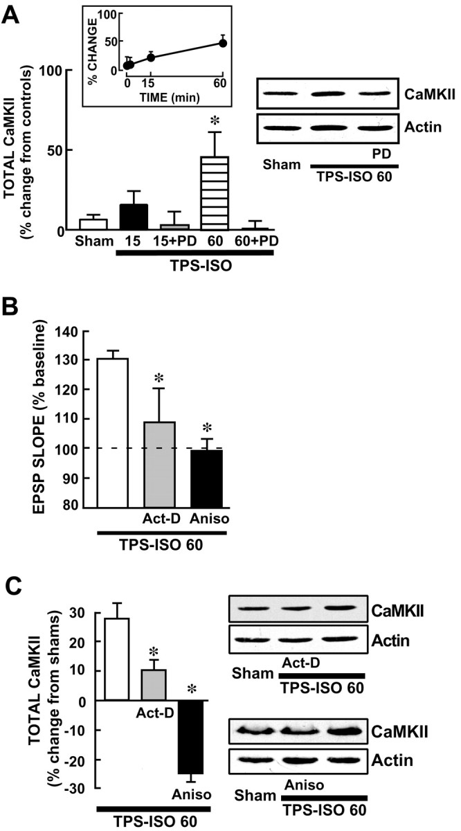
Fig. 6.

TPS–ISO produces a delayed increase in total CaMKII that requires MAPK activation and protein synthesis.A, A MEK inhibitor prevents the increase in CaMKII expression after TPS–ISO. Western immunoblots were prepared from CA1 regions, using antibody probes for total CaMKII and for actin. The bar graph shows summary data, with total CaMKII level normalized to actin from the same CA1 sample and expressed as percentage change from unstimulated controls. Total CaMKII was significantly increased only at 60 min after TPS–ISO (*p < 0.05). Atright is a representative immunoblot, which was run with CA1 homogenates from slices harvested at 60 min. PD= 50 μm PD98059. B, LTP measured at 60 min after TPS–ISO is inhibited by actinomycin-D or anisomycin. Both drugs were applied in the bath starting 30 min before TPS–ISO. The graph summarizes EPSP slopes at 60 min after stimulation, expressed as percentage of baseline. TPS–ISO-induced LTP measured 31.1 ± 6.7% above baseline (n = 5) and was blocked by 40 μm actinomycin-D (Act-D) or 20 μm anisomycin (Aniso) [+9.7 ± 11.7% (n = 3) and −1.1 ± 7.3% (n = 3), respectively; both pvalues < 0.05 vs TPS–ISO]. C, The increase in total CaMKII by TPS–ISO is blocked by actinomycin-D and anisomycin. Slices were harvested 60 min after TPS–ISO or at the equivalent time for sham-stimulated controls. The graph indicates total CaMKII levels normalized to actin for each band and expressed as percentage change from the sham-stimulated slices. TPS–ISO produced an increase in total CaMKII (+33.9 ± 16.6%; n = 5), which was prevented by 40 μm actinomycin-D and 20 μmanisomycin [+9.7 ± 6% (n = 3) and −24.2 ± 5.7% (n = 3), respectively; both pvalues < 0.05 vs TPS–ISO]. Anisomycin reduced total CaMKII below sham-stimulated controls (p < 0.05). Sample immunoblots are shown for CA1 homogenates from slices treated with actinomycin-D (top blot) and anisomycin (bottom blot).
