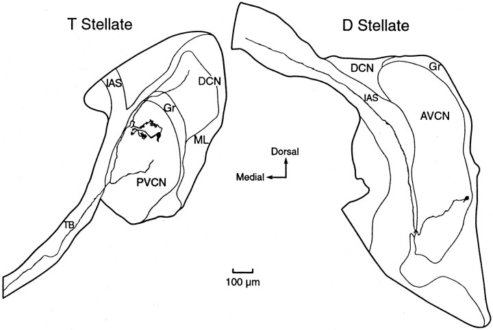
Fig. 2.
Reconstructions of biocytin-labeled T and D stellate cells of the VCN. Left, A T stellate cell whose responses to injected current are shown in Figure1A has a branched dendrite with a curly appearance. The axon characteristically projects out of the VCN through the trapezoid body and has collateral branches in the dorsal and ventral cochlear nuclei. Presumably some dendrites had been cut in preparing the slice. Right, D stellate cell whose responses to injected current are shown in Figure1D. The axon characteristically runs dorsally through the intermediate acoustic stria. Presumably the dendrites had been cut in the preparation of the slice. PVCN, Posteroventral cochlear nucleus; AVCN, anteroventral cochlear nucleus; DCN, dorsal cochlear nucleus;Gr, granule cell region; ML, molecular layer of DCN; TB, trapezoid body; IAS, intermediate acoustic stria.

