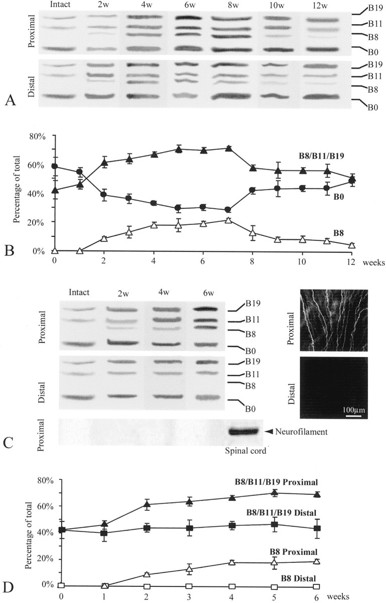
Fig. 2.

Upregulation of active agrin isoforms in adult Schwann cells induced by nerve regeneration. Expression of agrin isoforms in the Schwann cells along the frog sciatic nerve after short-term denervation (A, B) and long-term denervation (C, D) was examined by RT-PCR. A, Schwann cells in the intact sciatic nerve expressed only three agrin isoforms: B0, B11, and B19. However, after a single nerve transection that allowed nerve regeneration, Schwann cells in both the proximal and the distal nerve stumps upregulated the expression of active isoforms, including the appearance of B8. The number of weeks after axotomy is denoted on the top of each lane. B, A plot shows changes in the percentage of B0 (filled circles), B8 (open triangles), and all active isoforms (B8/B11/B19;filled triangles) relative to the total isoforms (see Materials and Methods) at different time points after short-term denervation (n = 3 experiments; mean ± SEM). The increase in the relative expression of active agrin and a concomitant decrease in B0 began at ∼2 weeks and peaked at ∼6–7 weeks after short-term denervation. A trace amount of B8 (3%) was still detected 12 weeks after axotomy. C, Schwann cells in the sciatic nerve segment proximal to the transection site 2–6 weeks after long-term denervation upregulated the active agrin isoforms compared with the intact nerve (top left panel). The same Schwann cell samples along the intact nerve and the proximal nerve segment 2–6 weeks after axotomy did not show any neurofilament mRNA, in contrast to the spinal cord (bottom left panel). The proximal segment contained regenerating axons, as revealed by positive anti-neurofilament staining (top right panel). In contrast, the distal segment, which was chronically severed from the proximal segment, was absent of anti-neurofilament staining (bottom right panel). Schwann cells in the distal segment showed neither upregulation of B11 and B19 nor appearance of B8 (middle left panel).D, A plot shows an increase in the relative expression of all agrin isoforms (B8/B11/B19;filled triangles) and B8 (open triangles) in the proximal segment after long-term denervation. However, in the chronically segregated distal segment, the relative expression of the total active isoforms (B8/B11/B19; filled squares) remained unchanged, and no B8 (open squares) was detected up to 6 weeks after axotomy (n = 3 experiments; mean ± SEM).
