Fig. 3.

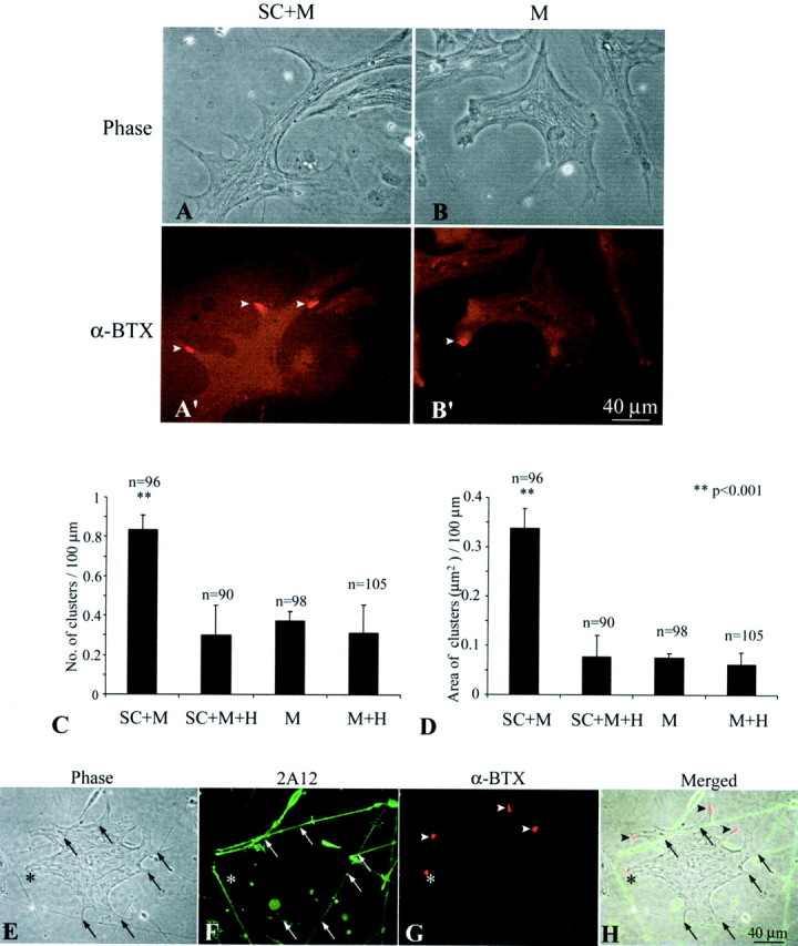
Adult Schwann cells enhance AChR aggregation on muscle in culture. Aggregation of AChRs on embryonicXenopus SC+M cells for 7 d was compared with that in M. A, B, Phase-contrast images of an SC+M culture (A) and an M culture (B) show similar muscle morphology.A′, B′, Fluorescence images of the same cultures labeled with Texas Red-conjugated α-BTX show more AChR hotspots in SC+M than M. C, A plot shows a significant increase in the number of AChR hotspots per 100 μm muscle length in SC+M, which was 2.2-fold of that in M. Treatment with heparin (300 μg/ml) eliminated this increase in the coculture (SC+M+H), but the treatment did not show effect in pure muscle culture (M+H).D, A plot shows the total area (in square micrometers) of AChR clusters per 100 μm of muscle length in SC+M, M, and after heparin treatment in SC+M+H and M+H. Similar to C, the area of AChR clusters in SC+M was significantly enlarged to 4.5-fold of that in M, and this increase was eliminated by heparin treatment. In C andD, n denotes the number of muscle fibers observed, and all values are mean ± SD. E–H, SC+M cocultures were examined to determine the spatial relationship between AChR aggregates and Schwann cell–muscle contacts. The contacts (arrows) could be observed with phase contrast optics (E) and confirmed with staining of Schwann cells with mAb 2A12 (F). The same culture double-labeled with α-BTX (G) showed that the majority of these contacts were not colocalized with AChR clusters (arrowheads). Only in rare cases were AChR clusters colocalized with the Schwann cell–muscle contact (asterisk). The spatial relationship between AChR clusters and the contacts is further shown in H, which is a merged image of E–G.
