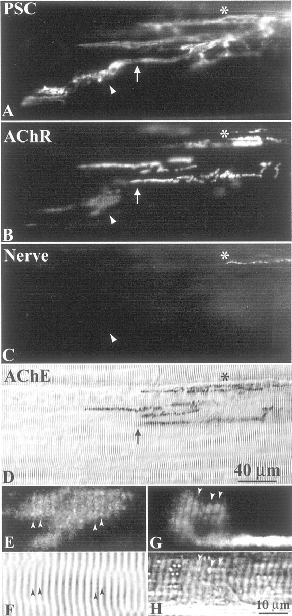
Fig. 6.

PSC sprouts may induce AChR aggregation in vivo. NMJs 4 weeks after axotomy were fluorescently labeled with mAb 2A12 for PSCs (A), α-BTX for AChRs (B), and antibodies to neurofilament 200 kDa and synapsin I for axons and nerve terminals, respectively (C). A, A prominent PSC sprout (arrowhead) extended beyond the boundary of the original junction delineated by AChE (arrow in D; corresponding arrows in A andB). B, A large extrajunctional AChR aggregate (arrowhead) colocalized with the PSC sprout (arrowhead in A). C, Axons and nerve terminals were absent from extrajunctional AChR aggregates (arrowhead) and PSC sprouts, suggesting that PSC sprouts may directly induce AChR clustering in vivo.Asterisk marks the closest extent of regenerating nerve terminals. D, The original synaptic sites were labeled with Karnovsky's AChE stain. The scale bar in D applies to A–D. E, A high-magnification view of the AChR aggregate marked by the arrowhead inB. F, Bright-field view of the same region of the muscle fiber as in E. The AChR aggregate showed sarcomeric staining pattern with stripes of brighter AChRs (arrowheads in E) matching with the light bands (arrowheads inF) of sarcomeres. G, An extrajunctional AChR aggregate in a reinnervated muscle (3 weeks after axotomy), which was freshly dissected and labeled with α-BTX without previous fixation, membrane permeabilization, or other staining procedures. H, Bright-field view of the same region of the muscle fiber as in G. The sarcomeric pattern also showed brighter stripes of AChRs (arrowheads inG) matching with the light bands (arrowheads in H) of sarcomeres. The scale bar in H applies to E–H.
