Fig. 3.
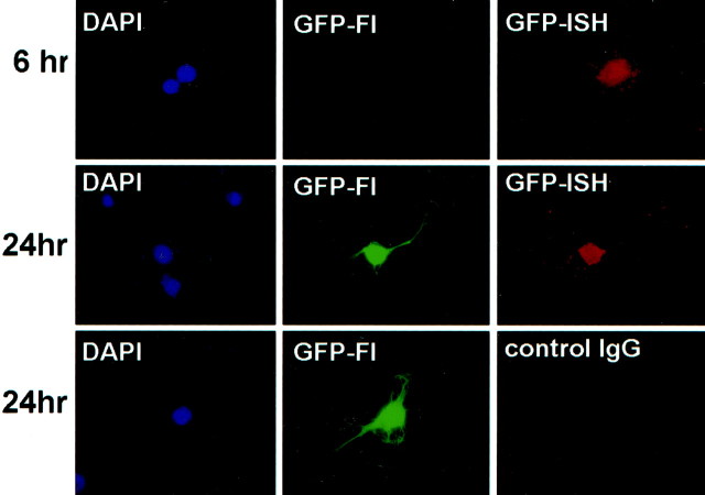
GFP mRNA and GFP protein expression in transfected neurons. Neurons were processed for GFP fluorescence (GFP-Fl) and fluorescent in situhybridization (GFP-ISH) at either 6 or 24 hr after transfection. Neurons at 6 hr can contain GFP mRNA without expressing detectable GFP fluorescence (top panel). In contrast, at 24 hr after transfection, all GFP mRNA-containing neurons also express the fluorescent GFP protein (middle panel). In the bottom panel, the anti-DIG primary antibody was replaced with a nonspecific normal mouse IgG antibody (control IgG). DAPI staining reveals the nuclei of cells within each field.

