
Fig. 7.
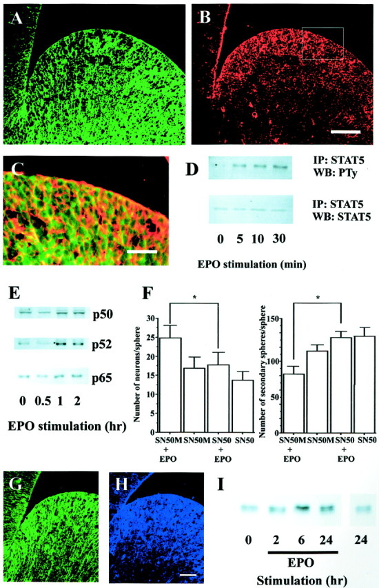
Putative signaling mechanisms for EPO actions on NSCs. A, B, EPO-R (A,red) and STAT5 (B, green) were coexpressed in the progenitor cell population of the E14 mouse ganglionic eminence, as shown by theorange–yellow staining in the merged image (C). The enlarged area in Cis that indicated by the rectangle in B. Scale bars: (shown in B for A,B), 100 μm; C, 50 μm. D, Passage 1 neurospheres from the E14 mouse ganglionic eminence were stimulated with 10 IU/ml EPO for the time periods indicated. Cellular extracts were immunoprecipitated (IP) with anti-STAT5, and immunoprecipitates were analyzed by Western blot (WB) with antiphosphotyrosine (PTy). Equalized protein loading was controlled for with anti-STAT5.E, Passage 1 neurospheres generated from the E14 mouse ganglionic eminence were stimulated with 10 IU/ml EPO for the time periods indicated. Nuclear extracts were analyzed by Western blotting with anti-NF-κB p50, p52, and p65 antibodies. F, Before EPO stimulation, passage 1 cells (clonal density) were treated with 10 μg/ml of SN50 or SN50M for 24 hr, and the media was then changed to EGF (with or without EPO)-containing media. Seven days after treatment, the spheres were dissociated for sphere-forming assays or processed to assess neuron number (*p < 0.05).G, H, EPO-R (G,green) and Mash1 (H, blue) were coexpressed in the progenitor cell population of the E14 mouse ganglionic eminence. Scale bar, 50 μm. I, Passage 1 neurospheres from the E14 mouse ganglionic eminence were stimulated with 10 IU/ml EPO for the time periods indicated, and WBs were used to analyze cellular extracts for Mash1 expression.