
Fig. 2.
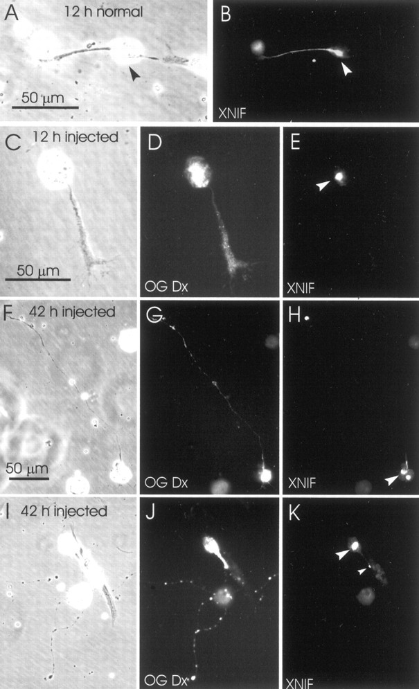
Effects of the injected anti-NF-M antibody on the intracellular distribution of XNIF. A, B, Typical distribution of XNIF immunoreactivity in a normal young neurite, 12 hr after plating. Staining extends from the cell body (arrowhead) through the neurite in a proximal-to-distal gradient of decreasing intensity. C–K, The distribution of XNIF in neurons descended from the blastomere that was injected with anti-NF-M. Left,center, and right columns show phase-contrast, Oregon Green Dextran 488 (OG Dx) fluorescence, and peripherin immunofluorescence views of the same neurons, respectively. C–E, Young neuron, 12 hr after plating. XNIF immunoreactivity was confined exclusively to the cell body (arrowhead).F–I, Older neurons, 42 hr after plating. XNIF immunoreactivity was still confined primarily to the cell body (large arrowheads) but also occasionally appeared as small dots scattered along the neurite (K, small arrowhead). The scale bars in A,C, and F also apply to B,D, E, andG–K, respectively.