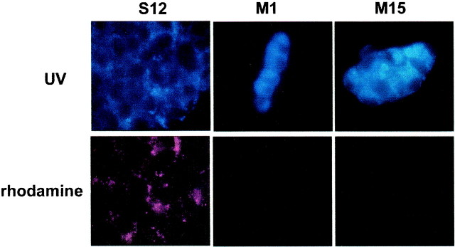
Fig. 10.
Reduced degradation of a lysosomal substrate in PC12 cells expressing A53T α-synuclein. PC12 cells from the various cell lines were incubated with 2 mg/ml Lysosensor Yellow/Blue Dextran for 20 hr, rinsed three times in complete medium, and then visualized under a 100× oil-immersion objective. Images were captured in a UV (top) or a rhodamine (bottom) filter. Note the increased fluorescent signal in cells from the M1 and M15 lines in the UV range, indicative of accumulation of the substrate in nonacidic organelles, and the absence of fluorescence in the rhodamine spectrum, indicative of the absence of degradation of the substrate in acidic organelles. Identical exposure times were used across the various lines. The experiment was repeated twice with similar results.

