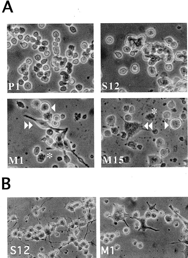
Fig. 2.

Morphological alterations in PC12 cells expressing A53T α-synuclein. A, Photomicrographs of naive PC12 cells expressing empty vector (P1), wild-type α-synuclein (S12), or A53T mutant α-synuclein (M1 and M15). Theasterisks indicate granular degenerating cells. Thesingle arrowhead indicates very large cells. Thedouble arrowhead indicates a neuritic-like extension in an M1 cell and a stellate appearance in a M15 cell. B, Photomicrographs of PC12 cells expressing wild-type α-synuclein (S12) or A53T mutant α-synuclein (M1) and treated for 9 d with NGF. Note the neuritic network present in the S12 cultures but not in the M1 cultures. Also note the large number of degenerating cells in the cultures derived from the M1 line.
