
Fig. 7.
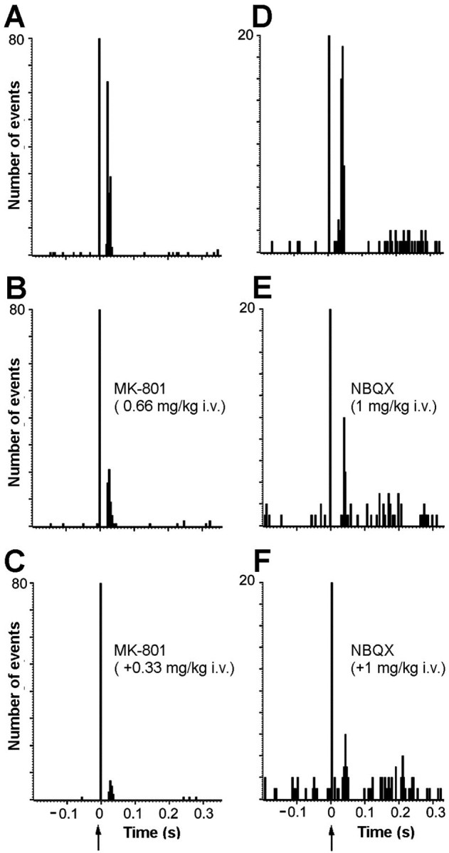
Involvement of ionotropic glutamate receptors in the mPFC-induced activations of DR 5-HT neurons. A shows the baseline orthodromic activation (52%, latency 18–36 msec) of a 5-HT neuron after the stimulation of mPFC. B andC show, in the same neuron, the reversal of the excitation produced by increasing doses of the NMDA receptor antagonist (±)MK-801 (0.66 + 0.33 mg/kg, i.v.). Bin size: 4 msec, 240 sweeps.D shows the baseline orthodromic activation (21.2%, latency 26–48 msec) of another DR 5-HT neuron after the stimulation of mPFC. E and F show the reversal of the excitation produced by increasing doses of the AMPA–KA receptor antagonist NBQX (1 + 1 mg/kg, i.v.) in the neuron shown inD. Bin size: 4 msec, 225 sweeps.