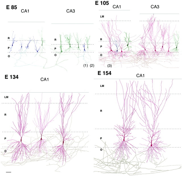
Fig. 1.
Morphological differentiation of pyramidal cells in the cynomolgus monkey hippocampus during the second half of gestation. Reconstruction of biocytin-filled pyramidal cells in CA1 and CA3 hippocampal subfields at various ages in utero(E85–E154). Note an intensive growth of the pyramidal cells that reach a high level of morphological differentiation at E134, 1 month before birth. O, Stratum oriens; P, pyramidal cell layer; R, stratum radiatum; LM,stratum lacunosum moleculare; the dashed lines indicate the limits between layers, and the solid line indicates the hippocampal fissure. Axons (gray), color of the dendritic arborization indicates the expression of synaptic currents (blue, silent; green, GABA-only;red, GABA + glutamate neurons). Electrophysiological recordings from neurons 1–3 are shown on Figure 3A. Scale bar, 100 μm.

