Fig. 5.
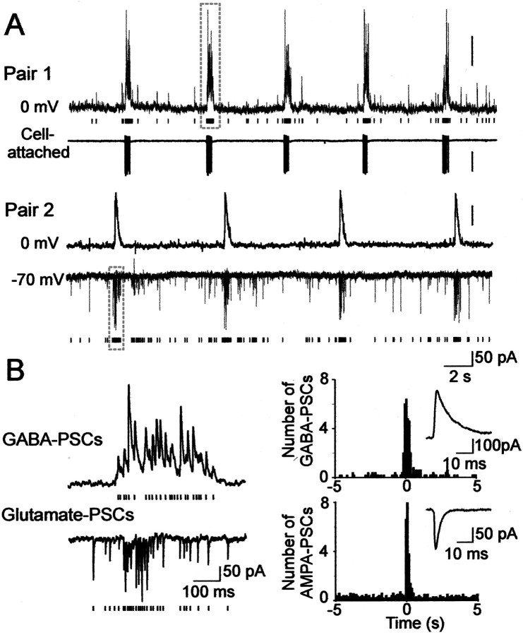
Giant depolarizing potentials synchronize most of the hippocampal neuronal activity in utero. A, Pair recordings of CA3 pyramidal cells and interneurons (E109). Pair 1, The pyramidal cell (top trace) is recorded in the whole-cell mode at the reversal potential of glutamatergic PSCs (0 mV), and the GABA(A) PSCs are outwardly directed; the hilar interneuron (bottom trace) is recorded in the cell-attached mode. Each GABA(A) PSC detected in the pyramidal cell is shown as abar below. Note the periodic oscillations (GDPs) synchronously generated in both neurons and associated with an increase of the GABA(A) PSC frequency in the pyramidal cell and bursts of action potentials in the interneuron. Pair 2, The pyramidal cell (top trace) is recorded in whole-cell mode at 0 mV, and the interneuron (bottom trace) is recorded in whole-cell mode at the reversal potential of the GABA(A) PSCs (−70 mV) so that the AMPA PSCs are inwardly directed. Each AMPA PSC detected in the interneuron is shown as a bar below.B,Left, GDPs outlined by dashed boxes on A are shown on expanded time scale. Note an increase in the frequency of the GABA(A) and AMPA PSCs during GDPs. On the right, the distribution of GABA and AMPA PSCs is cross-correlated with the population discharge (bin size, 100 msec). Insets, Averaged GABA and AMPA PSCs.

