
Fig. 2.
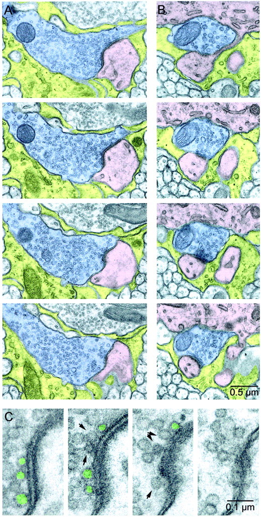
Serial EM sections of CFs. A,B, Two sample release sites from the same CF. CF axons are shaded blue, Purkinje cell dendrites and spines are shaded pink, and astrocytes are shadedyellow. The release site in A is larger than average, whereas the release site in B is smaller than average. In A, the synaptic cleft and PSD are clearly identifiable in the top three panels. InB, two spines are visible emerging from the parent dendrite at the top of each panel. The PSD for one spine is visible in the bottom three panels, and the second makes a PSD in later sections. C, Close-ups of the active zone in A. The presynaptic terminal is on the left, and the postsynaptic spine, with PSD clearly visible, is on the right. A vesicle was classified as docked (green) if it was located opposite a PSD and directly touched the presynaptic membrane. Nondocked vesicles close to the membrane are indicated byarrows.