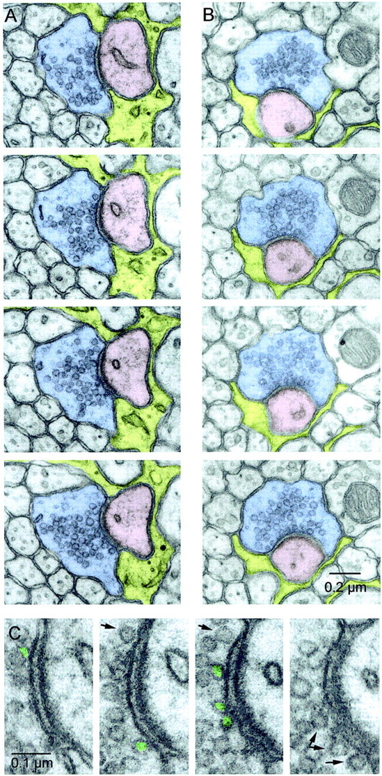
Fig. 5.

Serial EM sections of PFs. A,B, Two example series. PFs are shadedblue, Purkinje cell spines are shadedpink, and astrocytes are shaded yellow. Every panel contains a clearly defined synaptic cleft and PSD. The release site in A is larger than average, whereas the release site in B is smaller than average. Although neither PF has a mitochondrion in the sections shown here, the release site in A has a mitochondrion in a section that is 1.5 μm away, and the site in B has one in a section that is 0.2 μm away. C, Close-up of the active zone inA. The presynaptic terminal is on theleft, and the postsynaptic spine, with PSD clearly visible, is on the right. Docked vesicles are labeled ingreen, and nondocked vesicles close to the membrane are indicated by arrows.
