
Fig. 1.
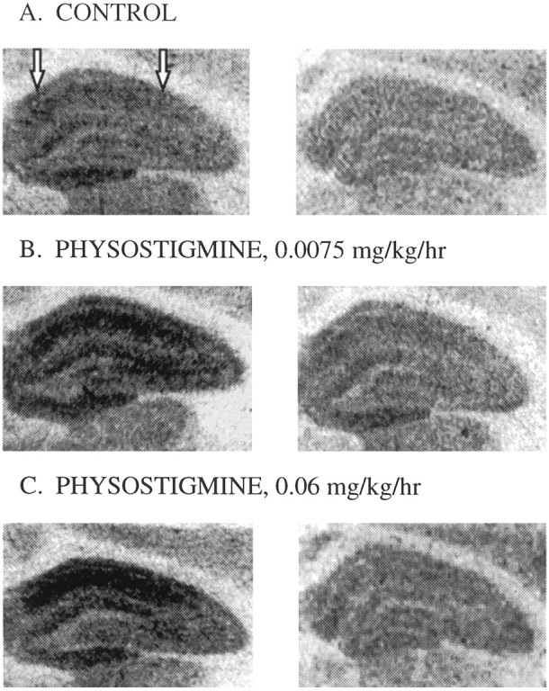
Representative autoradiograms of [3H]glutamate binding in the dorsal hippocampus in brains taken from ovariectomized rats receiving control treatment (A), 3 d of chronic treatment of physostigmine at 0.0075 mg · kg−1 · hr−1(B), or 3 d of chronic treatment of physostigmine at 0.06 mg · kg−1 · hr−1(C). Left panels illustrate total [3H]glutamate binding, and right panels illustrate [3H]glutamate binding that remained in the presence of NMDA. NMDA binding was taken to be the amount of total [3H]glutamate binding displaced by NMDA (difference between left and right panels). Arrows indicate the area in which measurements were taken.