
Fig. 8.
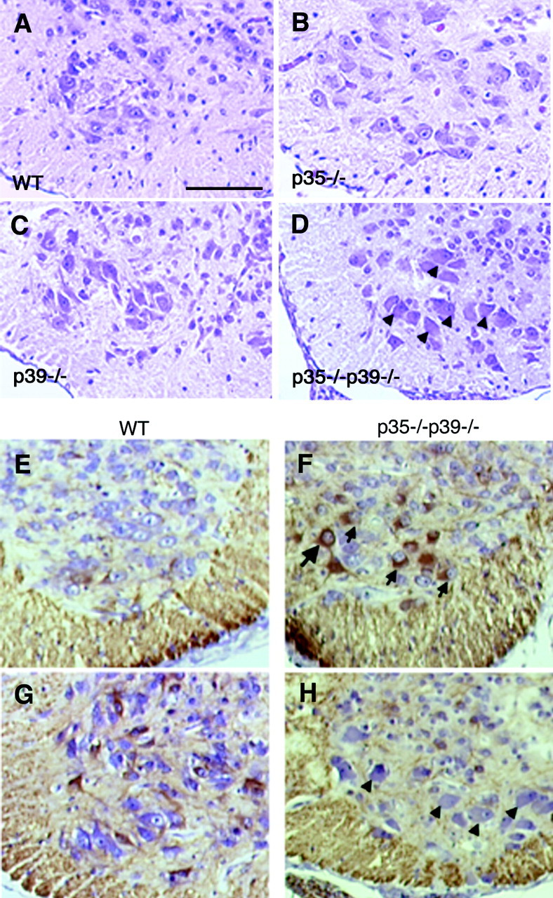
p35–/–p39–/– mice display motor neuron pathology. A, Transverse section through the lower thoracic spinal cord of a wild-type E18.5 pup shows large motor neurons in the anterior horn. B, C, Motor neurons of p35–/– (B) and p39–/– (C) mice appear primarily normal. D, In p35–/–p39–/– mice, the motor neurons exhibit ballooned perikarya and eccentric nuclei, which are hallmarks of chromatolysis. These chromatolytic changes are also seen in Cdk5–/– mice (reported previously).E–H, Immunostaining transverse sections of the wild-type (E, G) and p35–/–p39–/– (F, H) lower cervical (E, F) and lower thoracic spinal cord (G, H) with SMI34, a monoclonal antibody that detects a phosphorylated epitope of NF-H, shows that motor neurons of p35–/–p39–/– mice have aberrant accumulation in the soma of phosphorylated NF-H (F, arrows) that is normally found in axon fibers (E, G). SMI34-positive motor neurons, however, do not always display ballooned perikarya (F,large arrow). Conversely, ballooned neurons are not consistently labeled by SMI34 (H,arrowheads). This may reflect different stages in pathology and related changes in neurofilament phosphorylation. Scale bar, 150 μm.