
Fig. 10.
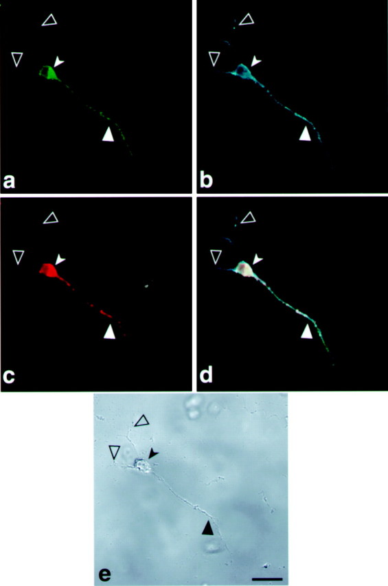
Tau-targeting signal drives MAP2 expression into the axon of differentiated P19 cells. Confocal image of a P19 cell line transfected with a construct containing GFP-MAP2-cod-fragment-H of tau 3′UTR. a, Localization of GFP-MAP2 protein in the axons.b, P19 cell stained with MAP2 antibodies shows the dendrites and axon. c, Localization by in situ hybridization of GFP-MAP2 mRNA with a GFP probe detected with anti-dig HRP/Cy5. d, Merged image ofa, b, and c showing the colocalization of GFP-MAP2 protein and mRNA in the axon.e, Phase view. Scale bar, 10 μm. Large filled arrowheads denote an axon, large open arrowheadsdenote dendrites, and small solid arrowheads denote a neuronal cell body.