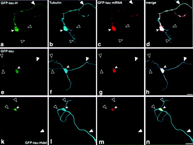
Fig. 4.
Confocal microscopy image of P19 cell cultures analyzed by in situ hybridization combined with immunohistochemistry using tubulin antibodies. a–d, Confocal image of a P19 cell line transfected with GFP-tau–cod-H construct. e–h, Confocal image of a P19 cell line transfected with GFP-tau–cod construct. i–l, Confocal image of a P19 cell line transfected with GFP-tau–cod-Hdel construct. a, e, i, GFP-tau fluorescence. b, f,j, P19 cell immunostained with tubulin antibodies.c, g, k, In situ hybridization using a GFP probe labeled with UTP-dig and detected with anti-dig HRP/Cy5. d, h,l, Merged confocal image showing colocalization of GFP-tau fluorescence with tubulin and with GFP-tau mRNA. Scale bars, 10 μm. Large filled arrowheads denote an axon,large open arrowheads denote dendrites, and small filled arrowheads denote a neuronal cell body.

