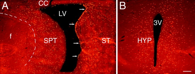
Fig. 2.
The distribution of newly generated cells in the parenchyma surrounding the lateral and third ventricles after the coinfusion of BDNF and BrdU into the lateral ventricle of an adult rat brain. The newly generated cells (bright orange) are identified in 20 μm coronal sections with an antibody to BrdU and visualized with a rhodamine-conjugated secondary antibody.A, A representative fluorescent photomicrograph demonstrating BrdU+ cells in the parenchyma surrounding the infused lateral ventricle 16 d after a 12 d infusion of BDNF–BrdU. The striatal SVZ (arrows) has numerous BrdU+ cells, whereas the rest of the SVZ, including that lining the septum, is almost devoid of newly generated cells. Moreover, the dorsal half of the striatal SVZ appears thicker than the ventral part. The distribution of the BrdU+cells in the striatal parenchyma exhibits a medial to lateral gradient, with the number of BrdU+ cells decreasing as a function of distance from the lateral ventricle. The distribution of BrdU+ cells in the septal parenchyma is more homogenous, although there is a relatively sharp decrease in the number of BrdU+ cells at the border between septum and fornix (dashed line). Note that, on both sides of the lateral ventricle, the BrdU+ cells extend more than a few hundred micrometers into the parenchyma. A small number of the newly generated cells can also be observed in the corpus callosum overlying the lateral ventricle. The midline of the section is approximately at the left edge of the photomicrograph.B, A representative fluorescent photomicrograph showing BrdU+ cells in the parenchyma surrounding the third ventricle of a BDNF-infused brain. In the hypothalamus, the newly generated cells extend bilaterally at least a few hundred micrometers into the parenchyma, and their distribution is relatively homogenous. Similar to the septal SVZ (shown in A), the hypothalamic ventricular lining is devoid of BrdU+cells. 3V, Third ventricle; CC, corpus callosum; HYP, hypothalamus; LV, lateral ventricle; SPT, septum; ST, striatum. Scale bar, 100 μm.

